Colorful Neurons
2016-08-22 |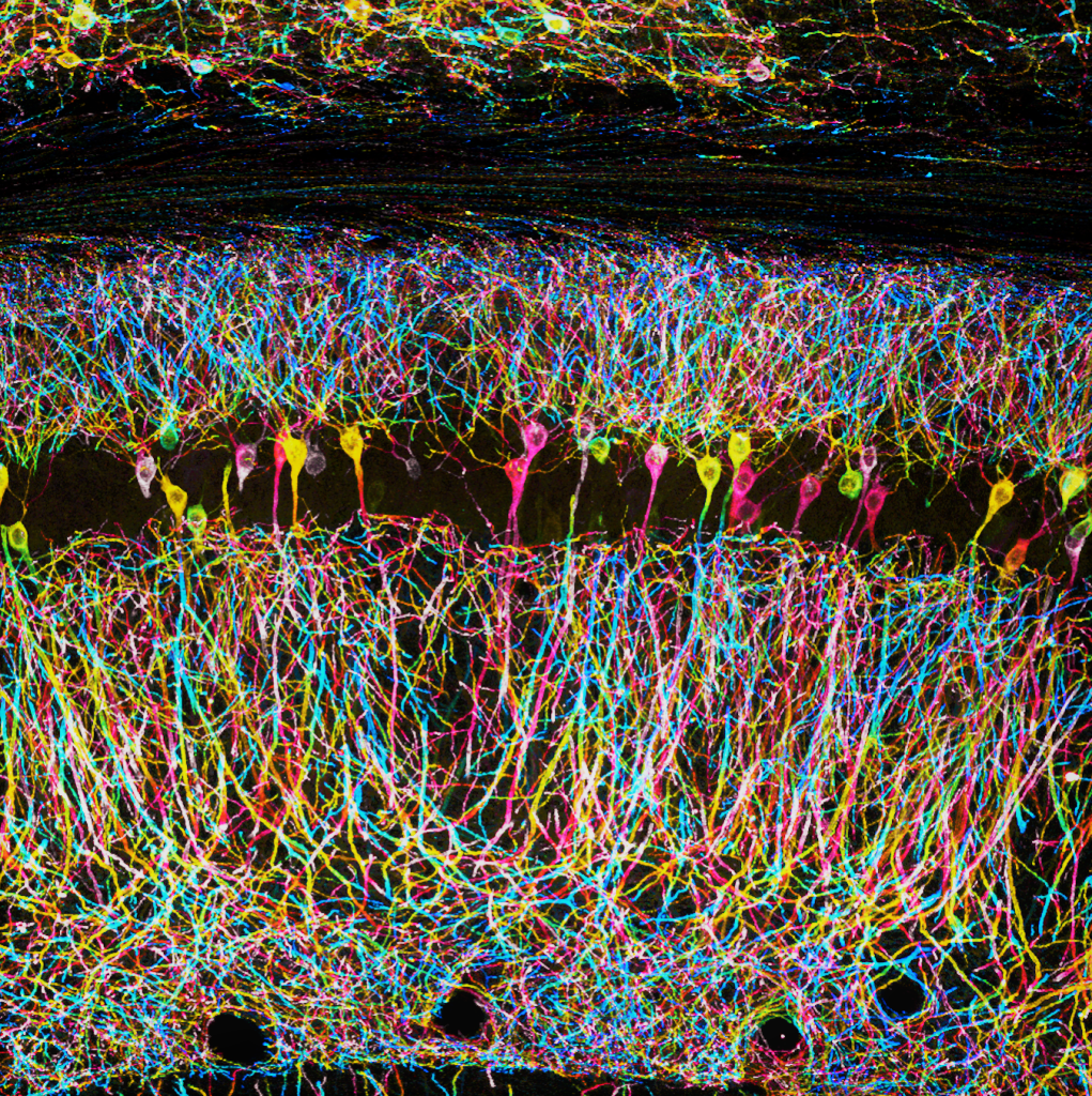
Neurons are not colorful. they appear grey/white-ish under a regular microscope.
However, if we selectively label a subset of these neurons with fluorescent proteins they will glow under the light (or laser) of a microscope.
How to acquire these images
The microscope takes thin optical slices (i.e. pictures) of these neurons along the Z axis (depth) allowing the observer to appreciate the 3D structure of the neurons.
Here I have projected the images (optical slice) along the Z-axis and assigned a different color to each optical slice using the beloved Fiji (ImageJ).
The result is a powerful image that reveals the complexity of the network (85-90% of the neurons in this picture are not visible!) and uses color in a clever way: neurons and neuronal processes lying on the same plane are labeled with the same color, while neurons and processes lying on different planes have different colors.